
Page 32 - MDJ 2022 Jan-Jun, Volume 45 Number 1
P. 32
Ahmad / Sebastian Rajah
coronal leakage may be an important cause of failure of treatments group. These root canal fillings showed
5
endodontic treatment. obturations that were short of radiographic apex, presence
of voids, and presence of periapical radiolucency. All these
There are also some cases in which the treatment failed root-treated teeth were recorded in a logbook
has followed the highest technical standards and yet prepared by this unit since 2009. A final sample of 165
resulted in failure. Scientific evidence indicates that some root-treated teeth were chosen after careful selection. Only
factors may be associated with the unsatisfactory outcome clearly visible radiographs which include the whole root
of well-treated cases, including extraradicular infection, length and at least 2 mm of the periapical region were
6–8
foreign body reactions, and true cysts. According to the selected. Poor quality radiographs as well as cases with
European Society of Endodontology, root canal treatment iatrogenic errors such as perforations, separated
9
has an unfavourable outcome when: instruments, and crown-root fractures were excluded. All
radiographs included were in the form of periapical films.
(1) the tooth is associated with signs and symptoms of
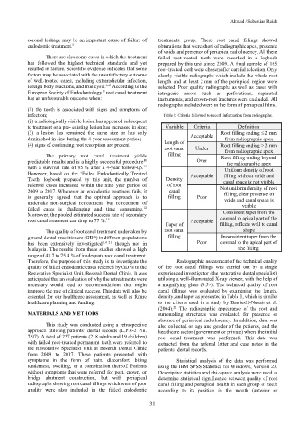
infection; Table 1: Criteria followed to record information from radiographs.
(2) a radiologically visible lesion has appeared subsequent
to treatment or a pre- existing lesion has increased in size; Variable Criteria Definition
(3) a lesion has remained the same size or has only Acceptable Root filling ending ≤ 2 mm
diminished in size during the 4-year assessment period; from radiographic apex
(4) signs of continuing root resorption are present. Length of Root filling ending > 2 mm
root canal Under from radiographic apex
The primary root canal treatment yields filling Root filling ending beyond
10
predictable results and is a highly successful procedure Over the radiographic apex
11
with a survival rate of 95 % after a 4-year follow-up. Uniform density of root
However, based on the ‘Failed Endodontically Treated Acceptable filling without voids and
Teeth’ logbook prepared by this unit, the number of Density canal space is not visible
referred cases increased within the nine year period of of root Not uniform density of root
2009 to 2017. Whenever an endodontic treatment fails, it canal
is generally agreed that the optimal approach is to filling Poor filling, clear presence of
voids and canal space is
undertake non-surgical retreatment, but retreatment of
12
failed cases is challenging and time consuming. visible
Moreover, the pooled estimated success rate of secondary Consistent taper from the
root canal treatment can drop to 77 %. Acceptable coronal to apical part of the
13
Taper of filling, reflects well to canal
The quality of root canal treatment undertaken by root canal shape
general dental practitioners (GDP) in different populations filling Inconsistent taper from the
has been extensively investigated, 14–21 though not in Poor coronal to the apical part of
Malaysia. The results from these studies showed a high the filling
range of 43.7 to 75.4 % of inadequate root canal treatment.
Therefore, the purpose of this study is to investigate the Radiographic assessment of the technical quality
quality of failed endodontic cases referred by GDPs to the of the root canal fillings was carried out by a single
Restorative Specialist Unit, Beserah Dental Clinic. It was experienced investigator (the restorative dental specialist)
anticipated that an evaluation of why the retreatments were utilising a well-illuminated X-ray viewer, with the help of
necessary would lead to recommendations that might a magnifying glass (3.5×). The technical quality of root
improve the rate of clinical success. This data will also be canal fillings was evaluated by examining the length,
essential for our healthcare assessment, as well as future density, and taper as presented in Table 1, which is similar
healthcare planning and funding. to the criteria used in a study by Barrieshi-Nusair et al.
(2004). The radiographic appearance of the root and
22
MATERIALS AND METHODS surrounding structures was evaluated for presence or
absence of periapical radiolucency. In addition, data was
This study was conducted using a retrospective also collected on age and gender of the patients, and the
approach utilising patients’ dental records (L.P.8-2 Pin. healthcare sector (government or private) where the initial
7/97). A total of 237 patients (218 adults and 19 children) root canal treatment was performed. This data was
with failed root-treated permanent teeth were referred to extracted from the referral letter and case notes in the
the Restorative Specialist Unit at Beserah Dental Clinic patients’ dental records.
from 2009 to 2017. These patients presented with
symptoms in the form of pain, discomfort, biting Statistical analysis of the data was performed
tenderness, swelling, or a combination thereof. Patients using the IBM SPSS Statistics for Windows, Version 20.
without symptoms that were referred for post, crown, or Descriptive statistics and chi-square analysis were used to
bridge abutment construction, but with periapical determine statistical significance between quality of root
radiographs showing root canal fillings which were of poor canal filling and periapical health in each group of teeth
quality were also included in the failed endodontic according to its position in the mouth (anterior or
31