
Page 33 - MDJ 2022 Jan-Jun, Volume 45 Number 1
P. 33
Radiographic Assessment of the Technical Quality of Failed Endodontic Treatment Referred from General Dental Practitioners in
Pahang, Malaysia: A retrospective study
posterior). A p value equal to or less than 0.05 was fillings, 80.0 % were inadequate (Table 5). Maxillary
considered statistically significant. incisors had the highest frequency of inadequate root
fillings at 49.2 %, followed by mandibular molars
Table 2: Distribution of root treated teeth in both jaws. (21.2 %), and maxillary premolars (12.9 %). Adequacy of
root canal fillings was also assessed in relation to tooth
Tooth type n % group. There was no statistically significant difference (p >
Maxillary incisors 88 53.3 0.05) in quality of root canal fillings among the different
Maxillary canines 1 0.6 tooth groups.
Maxillary premolars 18 10.9
Maxillary molars 9 5.5 Table 3: Distribution of root-treated teeth in relation to tooth
Total maxillary teeth 116 70.3 position.
Mandibular incisors 3 1.8 Tooth position n %
Mandibular canines 1 0.6 Maxillary anterior 8 9 5 3 . 9
Mandibular premolars 13 7.9 Maxillary posterior 2 7 1 6 . 4
Mandibular molars 32 19.4 Mandibular anterior 4 2 . 4
Total mandibular teeth 49 29.7 Mandibular posterior 4 5 2 7 . 3
RESULTS Healthcare Sector Where Initial
Endodontic Treatment Was
The subjects’ ages ranged from 11 to 71 years old, Performed
76 (46.1 %) were male and 89 (53.9 %) were female. The
distribution of the root-treated teeth is as tabulated in Table
2. The tooth most often referred for retreatment was the Missing
maxillary incisor (53.3 %), while maxillary and 9%
mandibular canines were the least referred (0.6 %). As a
group, more maxillary teeth were referred for retreatment
(70.3 %) compared to mandibular teeth (29.7 %). In
relation to tooth position, more maxillary anteriors (53.9 %) Private Govern
needed retreatment compared to mandibular anteriors 36% ment
(2.4 %) while more mandibular molars (27.3 %) needed 55%
retreatment compared to maxillary molars (16.4 %) (Table
3).
Symptoms in the form of pain, discomfort,
swelling, or a combination of symptoms were noted in 105
cases (63.6 %) while 60 cases (36.4 %) had no symptoms. Government Private Missing
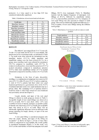
A total of 55.2 % patients had their initial root canal
treatment performed in a government clinic while 35.8 %
of patients had their initial root canal treatment done in a Figure 1: Healthcare sector where initial endodontic treatment
private clinic. The remaining 9.0 % of patients failed to was performed.
recall the sector in which their initial root canal treatment
was previously carried out (Figure 1).
Length, density, and taper of the root canal
fillings in relation to tooth group are shown in Table 4.
Acceptable length of root canal fillings was observed in
43.6 % of teeth, 54.5 % were short, while 1.8 % were
overfilled. In terms of density and taper, acceptable quality
of density was observed in 43.6 % of teeth and acceptable
taper was noted in 47.3 % of teeth. There was no
statistically significant difference between different tooth
groups regarding the length, density, or taper of the root
canal fillings.
A root canal filling is considered adequate only
when the length, density, and taper of the filling are
15
acceptable as shown in Figure 2. If one of these three
criteria is categorised as unacceptable, the overall quality
is considered inadequate (Figure 3). In this study, only Figure 2: Root canal filling 0–2 mm short of radiographic apex,
20.0 % of the total number of teeth had adequate root canal with uniform density and consistent taper (adequate).
32