
Page 42 - MDJ 2022 Jan-Jun, Volume 45 Number 1
P. 42
Said Gulam Khan / Khor / Mohd Sarmin / Jambai / Mohd Azlan / Nordin
DMSO. The sub-cultured bacteria were inoculated into concentration of the extract that showed no bacterial
BHI broth and the number of bacterial cells in the growth on agar medium was considered as the MBC. The
suspension were standardized to 0.08–0.13 using a test was done in triplicate.
8
spectrophotometer (OD 625 nm) which is equivalent to 10
cells/mℓ. Actively growing cultures were inoculated on a RESULTS
BHI agar plate. The disc with extract and controls were
placed aseptically on the inoculated medium. The plates AST of Ethanolic Extract of Z. mauritiana Leaves against
were incubated at 37 °C for 24–48 hours. The S. mutans
antimicrobial activity was evaluated by measuring the
diameter of the zones of inhibition, taken in three different The antibacterial activity of the extract was tested
directions. All tests were performed in triplicate. against S. mutans by disc diffusion method in the presence
14
of positive control (CHX) and negative control (20 %
Minimum Inhibitory Concentration (MIC) DMSO). The zone of inhibition was observed surrounding
the disc consisting of 200 and 500 mg/mℓ of Z. mauritiana
The antimicrobial activity of the ethanolic extract leaf ethanolic extract (Figure 4).
of Z. mauritiana leaves was further analysed using MIC
assay (CLSI, 2018). The concentration of the extract that Table 1: AST result of S. mutans against ethanolic extract
inhibits the growth of the bacteria from disc diffusion of Z. mauritiana leaves.
method was used for the MIC assay. Determination of
MIC of the extract was taken as the lowest concentration Zone of Inhibition (mm)
of the extract showing no visible growth of the organism Impregnated Disc Plate Plate Plate Mean
by standard micro-assays using sterile 96-well microplates. 1 2 3 (SD)
The sub-cultured bacteria were inoculated into BHI broth Positive CHX 27 26 26 26.33 ±
and the number of bacterial cells in the suspension was control 0.58
standardized to 0.08–0.13 using a spectrophotometer Negative 20 % 0 0 0 0 ± 0.0
8
(OD 625 nm) which is equivalent to 10 cells/mℓ. The control DMSO
standardized suspension was diluted to a ratio of 1:20 to 500 13.5 14.5 13.7 13.9 ±
yield 10 cells/mℓ. The plant extracts were serially diluted Z. mg/mℓ 0.53
6
in the 96-well with final concentration of extract ranging mauritiana 200 12 12 11 11.67 ±
from 200 to 0.1 mg/mℓ. The micro titre plate was labelled mg/mℓ 0.58
with number 1 until 12. 100 µℓ of BHI broth was pipetted
into all the wells (wells numbers 1 to 12). Then, 100 µℓ of
400 mg/mℓ of the plant extract was pipetted into well
number 1 as a starting concentration for MIC assay. Next,
a two-fold serial dilution was conducted by pipetting
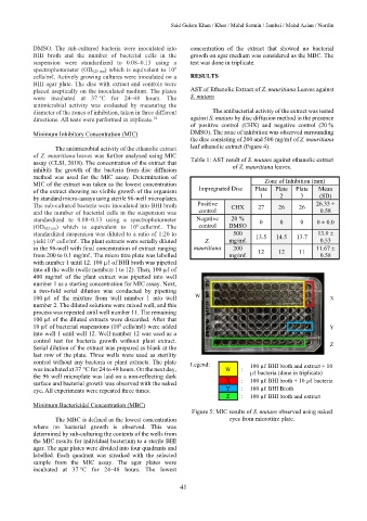
100 µℓ of the mixture from well number 1 into well W X
number 2. The diluted solutions were mixed well, and this
process was repeated until well number 11. The remaining
100 µℓ of the diluted extracts were discarded. After that
10 µℓ of bacterial suspensions (10 cells/mℓ) were added Y
8
into well 1 until well 12. Well number 12 was used as a
control test for bacteria growth without plant extract.
Serial dilution of the extract was prepared as blank at the Z
last row of the plate. Three wells were used as sterility
control without any bacteria or plant extracts. The plate Legend:
was incubated at 37 °C for 24 to 48 hours. On the next day, W : 100 µℓ BHI broth and extract + 10
the 96 well microplate was laid on a non-reflecting dark µℓ bacteria (done in triplicate)
surface and bacterial growth was observed with the naked X : 100 µℓ BHI broth + 10 µℓ bacteria
eye. All experiments were repeated three times. Y : 100 µℓ BHI Broth
Z : 100 µℓ BHI broth and extract
Minimum Bactericidal Concentration (MBC)
Figure 5: MIC results of S. mutans observed using naked
The MBC is defined as the lowest concentration eyes from microtitre plate.
where no bacterial growth is observed. This was
determined by sub-culturing the contents of the wells from
the MIC results for individual bacterium to a sterile BHI
agar. The agar plates were divided into four quadrants and
labelled. Each quadrant was streaked with the selected
sample from the MIC assay. The agar plates were
incubated at 37 °C for 24–48 hours. The lowest
41