
Page 10 - MDJ 2022 Jan-Jun, Volume 45 Number 1
P. 10
Wan Mustafa / Ng / Lim
0.5 millimetres per day for the remaining period until a
satisfactory result was obtained. The overjet was reduced
to about 4 millimetres at the end of the distraction period.
The total distraction time was 21 days.
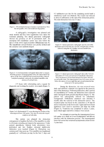
Figure 1: 3D reformatted image of patient at presentation. Note
the retrognathic chin and mandibular hypoplasia.
A radiographic investigation was planned and
study models and face bow registration were taken for
treatment planning. Her dental panoramic radiograph
showed the presence of all 32 permanent teeth. An
unerupted left mandibular canine was seen below the
apices of the lower right premolars (Mupparapu type 4).
The mandibular cortical borders were poorly defined and Figure 4: A dental panoramic radiograph near the end of the
the condyles were underdeveloped. distraction period showing the amount of lengthening of bone
achieved using the two Synthes extraoral multivector
distractors.
Figure 2: A dental panoramic radiograph taken prior to surgery.
Note the presence of transmigrated lower left canine below the Figure 5: A dental panoramic radiograph taken after forward
apices of the lower right first and second premolars, bilateral distraction of 16 mm and removal of mandibular distractors.
condylar hypoplasia, and poorly developed mandible with Note the presence of new bone between the lower first and
absence of cortical bone. second molar roots. The transmigrated lower left canine present
below the apices of lower right first and second premolars has
remained in the same position.
CT Scans with reformatting and a request for
diagnostic and working bio-models were made (Figure 3). During the distraction, she was reviewed twice
daily and antibiotic ointment was applied at the puncture
sites of the distractors. Following distraction, after a period
of consolidation of 7 weeks, the distractors were removed
under laryngeal mask general anaesthesia (Figure 5). The
patient was subsequently referred for orthodontic
management. Orthodontic treatment was initiated 8
months after the removal of the distraction device. The
treatment plan was based on the extraction of 14 and 24
and no extractions in the lower arch. Upper and lower fixed
appliances were used to reposition and align the teeth
(Figure 6). Treatment was completed in about 2 years,
upon which upper and lower retainers were issued.
Figure 3a–b: Reformatted CT scans showing the position of the
transmigrated lower left canine. Note also the underdeveloped
condyles on both sides. No attempts at removing the transmigrated lower
left canine were made as it was asymptomatic and did not
The patient was planned for distraction interfere with the distraction process. The transmigrated
osteogenesis to lengthen the body of the mandible in order lower left canine has remained asymptomatic at last review
to eliminate crowding and improve her appearance. Under 3 years later.
general anaesthesia, bilateral body osteotomies were
performed and two Synthes extraoral multivector
distractors were inserted (Figure 4). Distraction was
commenced 5 days post-op. The mandible was lengthened
gradually at 1 millimetre per day for the first 11 days and
9