
Page 11 - MDJ 2022 Jan-Jun, Volume 45 Number 1
P. 11
Transmigration of Mandibular Canine in a Patient with Hypoplastic Mandible: A case report
caused by lasting root formation, and a pericoronal
osteolytic area caused by widening of the follicular space.
In our case, the transmigration appears to be associated
1
with crowding secondary to mandibular hypoplasia.
Although the mandible was hypoplastic and therefore of
slightly reduced size, the canine was able to transmigrate
without hindrance and did not cause resorption of the roots
of the lower anterior and lower right premolar teeth. In our
patient, the mandibular hypoplasia affected the condylar
region more than the body. This meant that most of the
anterior mandibular height was retained, allowing
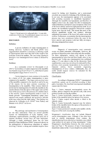
Figure 6: Dental panoramic radiograph taken 1.4 years after
completion of distraction. No pathological changes were seen in unhindered movement of the lower left canine across the
the transmigrated lower canine. midline. Furthermore, we postulate that the transmigration
process may have commenced before the roots of the
DISCUSSION lower anterior and premolar teeth had fully developed,
allowing unimpeded movement.
Definition
Diagnosis
A precise definition of canine transmigration is
7
lacking. However, Vuchkova and Farah (2010) have Diagnosis of transmigration most commonly
suggested there should be a consensus on defining a tooth occurs on dental panoramic radiographs. However, the
as transmigrated when more than half of the length of the problem of distortion exists when using only this modality.
tooth has crossed the midline. The case being reported is On these radiographs, canines may appear to have
therefore a true transmigrated lower canine as defined by transmigrated by more than half their length, when in fact
7
Javid. they have not. In this case, transmigration was confirmed
8
using a CT scan taken to plan for the distraction. Such
Incidence supplementary scans may not always be indicated but they
provide a more accurate interpretation on the position of
In a systematic review by Dalessandri et al. impacted canines, hence accurately diagnosing
(2017), the incidence of canine impaction in the mandible transmigration of lower canines. Additionally, they are
6
was found to range from 0.92 to 5.1 %, while that of canine useful should surgical removal be indicated for the
transmigration ranges from 0.1 to 0.31 %. transmigrated tooth.
Canine impaction is more common in the maxilla Classification
by a factor of 20, but canine transmigration is more
9
common in the mandible. The larger cross-sectional area According to Mupparapu (2002), transmigrated
13
of the mandible compared to the maxilla is said to be one mandibular canines can be classified according to their
of the reasons for the more common occurrence of path of deviation. He classified transmigrated mandibular
transmigration in the mandible. Other reasons which do canines into five types:
not favour the transmigration of teeth in the maxilla are the
smaller distance from the apices of the upper teeth and the Type 1: Canine impacted mesioangularly across the
floor of the nose as well as the presence of a midline suture midline, labial or lingual to the anterior teeth, with crown
in the maxilla. Canine transmigration tends to occur in portion of tooth crossing the midline.
7
female patients. Transmigrant mandibular canine cases Type 2: Canine horizontally impacted near the inferior
reported were usually unilateral, and involved a left-side border of the mandible below the apices of the incisors.
tooth more often than the right canine. This finding was Type 3: Canine erupted either mesial or distal to the
7
observed by Celikoglu et al. (2010) from Turkey and opposite canine.
10
Mazinis et al. (2012) in Greece. Type 4: Canine horizontally impacted near the inferior
11
border of the mandible below the apices of the premolar or
Aetiology molar on the opposite side.
Type 5: Canine positioned vertically in the middle with the
The aetiology and mechanism of transmigration long axis of the tooth crossing the midline.
are not fully understood. There are several possible
reasons for transmigration of teeth; tumours, cysts, Mupparapu’s classification does not include bilateral
odontomes, supernumeraries, crowding, and early loss or canine transmigration cases. Although it has often been
retained deciduous canines have all been implicated. used to classify transmigrant lower canines, the
Although a single aetiological factor has not been classification has been criticised as it includes cases which
7
12
identified, Pippi and Kaitsas (2008) proposed a theory on some authors do not agree as being transmigrant. Using
the pathogenesis of intraosseous migration of canines Mupparapu’s classification however, the most common
involving 2 factors: a strong and extended eruptive force type was Type 1. The presence of type 4 transmigrated
mandibular canine in this 13 year old girl is in contrast with
10