
Page 40 - MDJ Volume 47 Number 2 ( Jul-Dec 2024)
P. 40
Mustafa, et al.: Management of the Impacted Tooth with Dilacerated Roots – A Surgical Challenge
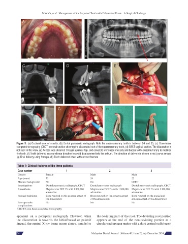
Figure 3: (a) Occlusal view of maxilla, (b) Dental panoramic radiograph. Note the supernumerary tooth in between 24 and 25, (c) Cone-beam
computed tomography (CBCT) coronal section showing the dilacerated root of the supernumerary tooth, (d) CBCT sagittal section. The dilaceration is
not seen in this view, (e) Access was obtained through a palatal flap, and elevators were used mesially and buccal to the supernumerary to mobilise
the tooth, (f) Tooth delivered in a curvilinear direction to avoid displacement into the antrum. The direction of delivery is shown in red (curve arrow),
(g) Final delivery using forceps, (h) Tooth delivered intact without root fracture
Table 1: Clinical features of the three patients
Case number 1 2 3
Gender Female Male Male
Age (years) 30 24 22
Medical background No No G6PD
Investigations Dental panoramic radiograph, CBCT Dental panoramic radiograph Dental panoramic radiograph, CBCT
Anaesthesia Mepivacaine HCl 2% with 1:100,000 Mepivacaine HCl 2% with 1:100,000 Mepivacaine HCl 2% with 1:100,000
adrenaline adrenaline adrenaline
Surgical technique Bone removal on the concave aspect of Bone removal on the concave aspect Bone removal on the mesial and
the dilaceration of the dilaceration concave aspect of the dilaceration
Post-operative No No No
complications
CBCT: Cone-beam computed tomography
apparent on a periapical radiograph. However, when the deviating part of the root. The deviating root portion
the dilaceration is towards the labial/buccal or palatal/ appears at the end of the non-deviating portion as a
lingual, the central X-ray beam passes almost parallel to circular radiopaque region with a dark central radiolucent
66 Malaysian Dental Journal ¦ Volume 47 ¦ Issue 2 ¦ July-December 2024
34