
Page 38 - MDJ Volume 47 Number 2 ( Jul-Dec 2024)
P. 38
Mustafa, et al.: Management of the Impacted Tooth with Dilacerated Roots – A Surgical Challenge
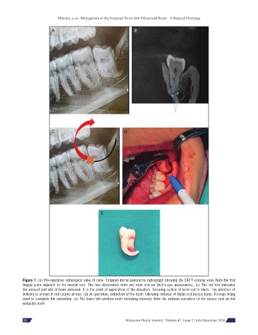
Figure 1: (a) Pre-operative radiological view of case. Cropped dental panoramic radiograph showing (b) CBCT coronal view. Note the thin
lingual plate adjacent to the mesial root. The two dilacerated roots are seen end on (bull’s-eye appearance), (c) The red line indicates
the amount and site of bone removed. X is the point of application of the elevators. Showing outline of bone cut in black. The direction of
delivery is shown in red (curve arrow). (d) At operation, extraction of the tooth following removal of distal and buccal bone. Forceps being
used to complete the extraction, (e) The lower left wisdom tooth following removal. Note the extreme curvature of the mesial root on the
extracted tooth
64 Malaysian Dental Journal ¦ Volume 47 ¦ Issue 2 ¦ July-December 2024
32