
Page 43 - MDJ Volume 47 Number 2 ( Jul-Dec 2024)
P. 43
Sharma, et al.: Gingivectomy via electrosurgery
orthodontic therapy. No significant past medical, family Intraoral examination revealed moderate chronic IGE
history or history of drug allergies were found. The on the marginal and papillary region of the gingiva. The
patient reported difficulty in maintaining oral hygiene provisional diagnosis of IGE induced by orthodontic
and experienced mild bleeding and discomfort. Clinical treatment was made.
examination confirmed IGE, likely exacerbated by
orthodontic appliances [Figure 1].
Management
Case presentation 2: Adult patient with moderate GE Phase 1 therapy was performed on all the patients on
A 36-year-old female patient reported with chief their first visit, and oral hygiene instructions were given
complaint of GE in the upper front tooth region for 6 along with a demonstration of the correct brushing
months after the beginning of orthodontic treatment. technique. The patients were then re-evaluated after 2
Medical and drug histories taken from the patient were weeks. Thereafter, the provisional diagnosis of chronic
negative. Intraoral examination revealed inflammatory IGE was made. After evaluating the extent of enlargement
enlargement was present in the marginal and papillary post non-surgical periodontal therapy, gingivectomy via
region of the gingiva, which was pink in colour, firm electrosurgery was chosen as the treatment modality to
and fibrotic in consistency, rounded interdental papilla, remove the excessive gingival tissue due to its precision
which was localised to six anterior teeth i.r.t #11–#13 and minimal bleeding. The patients were informed about
and #21–#23 [Figure 2]. Clinical examination provided the treatment, and written consent was taken from all
the provisional diagnosis of IGE induced by long-term three patients before starting the treatment procedure.
orthodontic treatment. Haematological examinations were done before starting
the surgical therapy to avoid any complications. Under
Case presentation 3: Teenager with localised GE aseptic conditions, local anaesthesia (2% lignocaine
with
hydrochloride
was
epinephrine)
1:80,000
A 19-year-old female patient reported the chief administered and pseudopockets were demarcated using
complaint of overgrown gingiva in her upper and a pocket marker [Figure 4]. An electrosurgical device was
lower front and back regions for a few months due used to remove excess gingival tissue and recontour of the
to prolonged orthodontic treatment [Figure 3]. The marginal gingiva during gingivectomy and gingivoplasty
enlargement caused aesthetic concerns and interfered procedures, respectively [Figure 5]. Universal curettes
with effective plaque control. No significant medical,
family history or history of drug allergies was found.
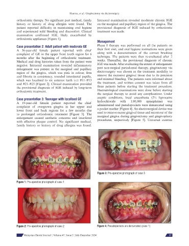
Figure 3: Pre-operative photograph of case 3
Figure 1: Pre-operative photograph of case 1
Figure 2: Pre-operative photograph of case 2 Figure 4: Pseudopockets are demarcated (case 1)
Malaysian Dental Journal ¦ Volume 47 ¦ Issue 2 ¦ July-December 2024 37