
Page 33 - MDJ Volume 47 Number 2 ( Jul-Dec 2024)
P. 33
Dhanrajani: Odontogenic keratocyst
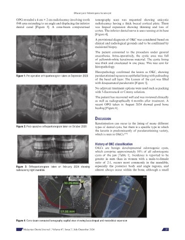
OPG revealed a 4 cm × 2 cm radiolucency involving tooth tomography scan was requested showing unicystic
#48 area extending to an angle and displacing the inferior radiolucency having a thick buccal cortical plate. There
dental canal [Figure 3]. A cone-beam computerised was lingual expansion showing thinning and loss of
cortex. The inferior dental nerve is seen running at its base
[Figure 4].
A provisional diagnosis of OKC was considered based on
clinical and radiological grounds and to be confirmed by
excisional biopsy.
The patient consented to the procedure under general
anaesthesia. Intra-operatively, the cystic area was full
of yellowish-white keratinous material. The cystic lining
was thick and enucleated in one piece. This was sent for
histopathology.
Histopathology confirmed the lesion as OKC, showing
Figure 1: Pre-operative orthopantomogram taken on September 2020 parakeratinised squamous epithelial lining with palisading
of the basal cell layer. The Lumen of the cyst was filled
with desquamated parakeratin [Figure 5].
No adjuvant treatment options were used such as packing
with 5-fluorouracil or Conroy solution.
The patient has recovered well and was reviewed clinically
as well as radiographically 6 months after treatment. A
recent OPG taken in August 2024 showed good bone
healing [Figure 6].
dIscussIon
Keratinisation can occur in the lining of many different
Figure 2: Post-operative orthopantomogram taken on October 2020 types of dental cysts, but there is a specific type in which
the keratin is predominantly of parakeratinising variety,
which is seen in OKCs. [5-7]
History of OKC classification
OKCs are benign developmental odontogenic cysts,
which comprise approximately 10% of all odontogenic
cysts of the jaw [Table 1]. Incidence is reported to be
greater in men than in women with a male-to-female
ratio of 2:1, occurs most commonly in the mandible,
Figure 3: Orthopantomogram taken on February 2024 showing especially the posterior body and angle regions, and
radiolucency right mandible almost always occur within the bone, although a small
Figure 4: Cone-beam computed tomography sagittal view showing buccolingual and mesiodistal expansion
Malaysian Dental Journal ¦ Volume 47 ¦ Issue 2 ¦ July-December 2024 27