
Page 34 - MDJ Volume 47 Number 2 ( Jul-Dec 2024)
P. 34
Dhanrajani: Odontogenic keratocyst
number of peripheral OKCs have been reported. [1,2] suggesting the term ‘Odontogenic Keratocyst’. Since
The term ‘Primordial cyst’ was first mentioned in 1945 then, OKC has been the most frequently researched cyst
by Robinson because the cysts were believed to have due to its high recurrence rate and aggressive clinical
primordial origin. In 1956, Philipsen and Reichart behaviour and associated with the nevoid basal cell
[1]
[2]
published a paper in Danish with an English summary carcinoma syndrome. [8,9]
In 2005 the WHO reclassified OKC as a neoplasm
and recommended keratocystic odontogenic tumour
(KCOT) as the appropriate designation. In justifying
the reclassification, they stressed ‘aggressive’ behaviour,
recurrence, the occasional occurrence of a ‘solid’ variant
and mutations in the protein patched homolog 1 (PTCH1)
gene.
The fourth edition of the WHO Classification of Head
and Neck Tumours, published in January 2017, has
reclassified odontogenic keratocystic tumour as OKC. [3-5]
OKCs are now considered benign cysts of odontogenic
origin. This has raised a debate between the groups who
considered OKCs as KCOT and vice versa. [2,3,5,6]
The fifth edition of the WHO classification of head
and neck tumours, published in January 2022, OKC
continues in the cyst classification and has the longest
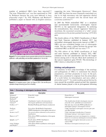
Figure 5: Photomicrograph showing parakeratinised stratified squamous
epithelium with palisading and abundant parakeratin in the lumen section among cysts of the jaw. Most show mutations of
the tumour suppressor gene PTCH1. The chronology of
OKC's history is described in Table 1.
Aetiology and pathogenesis
It is necessary to have some knowledge of the aetiology
and pathogenesis of OKCs to understand the clinical
presentation, and their propensity to recur. In the past,
OKCs were considered to originate from the primordium
of a tooth before its mineralisation and were called a
primordial cysts. [1,4-6] As understanding gained during the
Figure 6: Orthopantomogram taken on August 2024. Six months post- years passed the possibility gained ground that remnants
operative image showing good bone healing of dental lamina played a role in pathogenesis following
Table 1: Chronology of odontogenic keratocyst history
Author Description Main points
Robinson [1] Primordial origin Primordial cyst
Philipsen and Keratocyst Odontogenic keratocyst
Reichart6 [2]
Pindborg and Classification based on the interaction of This classification divided odontogenic tumours
Clausen [10] odontogenic epithelium and mesenchyme primarily in two: epithelial and mesenchymal
Gorlin et al. [11] Modified Pindborg and Clausen classification This had a key role in the WHO publication of
histological typing of odontogenic tumours
Pindborg First edition World Health Organisation Titled: histological odontogenic tumours, jaw cysts
et al. [12] (WHO) classification odontogenic tumour/cyst and allied lesions
Kramer et al. [13] The second edition elaborated on the previous Titled: histological classification of tumours:
classification histological typing of odontogenic tumours
Barnes et al. [14] Fifth edition WHO classification odontogenic Odontogenic keratocyst was designated as a tumour
tumour/cyst based on its recurrence odontogenic keratocyst (OKC)
Speight et al. [15] Fourth edition WHO Classification Reinstating odontogenic keratocyst as cyst, not
odontogenic tumour/cyst tumour OKC
Soluk-Tekkeşin Fifth edition WHO classification odontogenic Maintains odontogenic keratocyst as cyst
and Wright [16] tumour/cyst
60 Malaysian Dental Journal ¦ Volume 47 ¦ Issue 2 ¦ July-December 2024
28