
Page 28 - MDJ Volume 47 Number 2 ( Jul-Dec 2024)
P. 28
Fatima, et al.: Relationship of Cranial base growth with sagittal skeletal discrepancies
mean cranial base angles in patients with different skeletal In our study, mean posterior, anterior and total
sagittal discrepancies. In this study, the significance cranial base length was 46.10 ± 5.42, 65.69 ± 4.36 and
level was set at 5%, using the stratification technique 102.74 ± 7.84 mm, respectively. The mean NSAr and NSBa
the confounding factors such as age group and sex were cranial base angles were 127.25 ± 5.97° and 133.45 ± 6.34°.
modified and controlled. Post-stratification ANOVA test Detailed descriptive statistics are presented in Table 2
was applied.
Mean compassion of posterior, anterior and total cranial
base length and NSAr, NSBa cranial base angles according
to gender, age group and skeletal sagittal discrepancies
results
class was done and presented in Tables 3–8, respectively.
A total of 93 patients of either gender with ages ranging
from 13 to 30 years meeting inclusion criteria of the
study were evaluated to determine mean total, anterior dIscussIon
and posterior cranial base lengths and mean cranial Description and diagnosis of malocclusion is the
base angles amongst patients undergoing orthodontic primary objective of the orthodontist. The diagnosis
treatment as well as to compare the mean total, anterior can dictate the treatment objectives and treatment
and posterior cranial base lengths and mean cranial mechanics for the patient. Therefore, it is important to
base angles in patients with different skeletal sagittal find out if an underlying skeletal dysplasia is associated
discrepancies. with dental malocclusion. The location and magnitude
of skeletal dysplasia can influence various treatment
Amongst 93 patients, there were 25 males and 68 females. decisions. [9]
Out of 93 patients, 54.8% were from urban and 45.2%
were from rural areas, whereas 40.9% of patients were Debate has arisen in the selection of cranial base
classified as class I, 53.8% of patients were classified landmarks, over the use of the basion (Ba) or the articulare
[12]
as class II and 5.4% with class III. Detailed frequency (Ar). Bjork advocated the use of (Ar) point because it
distribution of gender, residential area and skeletal is easier and can better represent lateral cephalometric
sagittal discrepancies class are presented in Table 1. radiographs. However, Varjanne and Koski have suggested
the use of Basion despite the potential difficulties in
The overall mean age was 18.65 ± 3.16 years. The detailed identification because of its anatomic significance and
descriptive statistics of age are presented in Table 2. The discouraged the use of Articulare because of its remoteness
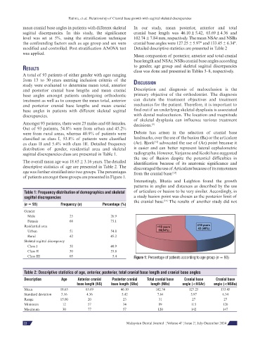
age was further stratified into two groups. The percentages from the cranial base. [13]
of patients amongst these groups are presented in Figure 1.
Interestingly, Bhatia and Leighton found the growth
patterns in angles and distances as described by the use
Table 1: Frequency distribution of demographics and skeletal of articulare or basion to be very similar. Accordingly, in
sagittal discrepancies a study basion point was chosen as the posterior limit of
the cranial base. The results of another study did not
[14]
(n = 93) Frequency (n) Percentage (%)
Gender
Male 25 26.9
Female 68 73.1
Residential area
Urban 51 54.8
Rural 42 45.2
Skeletal sagittal discrepancy
Class I 38 40.9
Class II 50 53.8
Class III 05 5.4 Figure 1: Percentage of patients according to age group (n = 93)
Table 2: Descriptive statistics of age, anterior, posterior, total cranial base length and cranial base angles
Description Age Anterior cranial Posterior cranial Total cranial base Cranial base Cranial base
base length (NS) base length (SBa) length (NBa) angle (<NSAr) angle (<NSBa)
Mean 18.65 65.69 46.10 102.74 127.25 133.45
Standard deviation 3.16 4.36 5.42 7.84 5.97 6.34
Range 15.00 20 23 31 27 27
Minimum 12 57 34 89 115 120
Maximum 30 77 57 120 142 147
54 Malaysian Dental Journal ¦ Volume 47 ¦ Issue 2 ¦ July-December 2024
22